DISCUSSION
There are numerous indications for root coverage procedures including sensitivity and esthetics. Esthetics may range from recession on a natural tooth to a newly visible margin on a porcelain fused to metal crown. The semilunar coronally advanced flap with the utilization of tricalcium phosphate has numerous advantages over other techniques for root coverage. First, by utilizing a conservative incision and maintaining the integrity of the interproximal papillae, the blood supply is preserved. This significantly improves healing time and overall success of the procedure. Furthermore, this technique does not require the use of sutures, which has multiple advantages: it minimizes tissue trauma, maintains the integrity of the blood supply, and allows for faster healing. The utilization of sutures in soft tissue augmentation procedures has multiple negative side effects, such as post-operative tissue shrinkage and acting as a reservoir for bacteria and food. This induces further inflammation of the tissue and causes delayed healing. The additional use of tricalcium phosphate has numerous benefits. Carranza et al. explains that calcium phosphate materials are biocompatible and osteoconductive.1 As demonstrated in this technique, tricalcium phosphate establishes hemostasis and functions as a scaffold for wound healing.1 One of the overarching goals of soft tissue surgeries is to regenerate the lost periodontal attachment apparatus; many studies have demonstrated the usefulness of tricalcium phosphate in these techniques. McGuire et al. compared the use of tricalcium phosphate and recombinant human platelet derived-growth factors with a coronally advanced flap to the traditional subepithelial connective tissue graft.(4) While both techniques demonstrated clinical success in achieving root coverage, the growth-factor mediated approach histologically demonstrated formation of cementum, connective tissue fibers, and supporting alveolar bone.(4) The tricalcium phosphate dually functions as a graft and membrane, promotes the establishment of hemostasis, and leads to the development of keratinized tissue.
While this procedure has the best prognosis in cases that present with Miller3 Class I defects and select Class II defects, it can also be performed in patients with more extensive recession. In more severe cases, the procedure can be done multiple times, each time gaining approximately 1-2 mm of gingival coverage until the desired gingival margin is achieved. It is important to note that this technique can be performed on a single tooth or two adjacent teeth simultaneously. Achieving root coverage on adjacent teeth using other techniques becomes more difficult due to donor site morbidity, lack of blood supply, and predictability.
Patient selection is crucial to the success of this procedure. Patients must demonstrate dili.gent oral hygiene compliance to ensure a predictable outcome. Additionally, this technique is well accepted by patients and is a practice builder for many reasons. The procedure can be performed in approximately fifteen minutes and patients generally do not require analgesics or antibiotics. As there is minimum postoperative discomfort, patients can engage in their daily activities with no impediment.


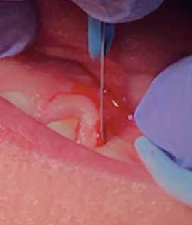 Gingival recession and inadequate keratinized tissue are prevalent problems encountered in dental practice and negatively impact prosthodontic and esthetic outcomes. Traditionally, gingival recession has been treated with numerous types of soft tis.sue procedures, which include coronally positioned flaps, connective tissue grafts, lateral and pedicle grafts, as well as guided tissue regeneration. These procedures are often lengthy, induce donor morbidity, and have poor patient acceptance because of post-operative pain. The semi-lunar incision technique was introduced by Dr. Dennis Tarnow in 1985. The technique discussed in this article builds upon the basic principles of the semilunar incision technique with the introduction of tricalcium phosphate, has a successful prognosis, and is well accepted by patients.

